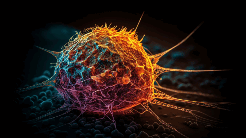
LA-ICP-TOFMS Imaging Reveals Significant Influence of Cancer Cell Resistance on Oxaliplatin Compartmentalization in the Tumor Microenvironment
Schaier et al.
JACS Au, 2025
DOI: 10.1021/jacsau.5c00217
최근 발표된 JACS AU by researchers from the 비엔나 대학교 및 Medical University of Vienna used an icpTOF 2R for single-cell analysis. The study found that oxaliplatin-resistant colorectal tumors altered drug distribution, with platinum accumulating in necrotic regions. Structural differences and resistance-dependent drug compartmentalization highlight how intrinsic cancer drug resistance affects therapeutic efficacy within the tumor microenvironment.
Colorectal cancer (CRC) treatment faces major setbacks due to oxaliplatin (OxPt) resistance, which can arise from complex cancer cell-intrinsic mechanisms and interactions within the tumor microenvironment (TME). While OxPt is widely used in both adjuvant and neoadjuvant settings, resistance, either intrinsic or acquired, remains a major challenge. This study investigates how OxPt-resistant CRC cells influence drug distribution in the TME using xenografts from HCT116 and OxPt-resistant HCT116/OxR cells. A novel analytical pipeline combining laser ablation inductively coupled plasma time-of-flight mass spectrometry (LA-ICP-TOFMS) and metal-labeled antibody-based multiplexed immunohistochemistry enabled single-cell level platinum mapping in tumor sections.
Using LA-ICP-TOFMS, the study achieved high-resolution, multiplexed imaging of oxaliplatin (OxPt) distribution in HCT116 and HCT116/OxR tumors at the single-cell level. Despite similar overall Pt levels in both models, OxPt accumulated primarily in stromal and necrotic regions, with minimal presence in proliferating cancer cells. Notably, resistant HCT116/OxR tumors exhibited higher Pt levels in necrotic zones and cancer-associated fibroblasts (CAFs), suggesting sequestration away from viable cells. Single-cell analysis using PhenoGraph and marker co-staining revealed that epithelial cells retained low Pt levels, while CAFs and necrotic cells harbored the highest. The MeXpose pipeline enabled sensitive Pt quantification (LOD: 0.23 fg/cell). Systemically, the spleen showed the highest Pt accumulation, suggesting hematotoxicity.
These results highlight how spatially resolved LA-ICP-TOFMS profiling reveals resistance-associated drug distribution patterns that impact OxPt efficacy. Cancer drug resistance alters both TME structure and platinum localization, with resistant tumors showing Pt accumulation in necrotic regions. These results suggest that resistant tumor cells can reprogram the TME to support therapeutic failure, highlighting the importance of spatially resolved, high-resolution analysis using an icpTOF 2R in understanding chemoresistance.
